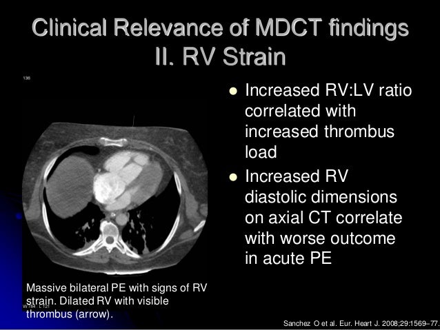
Rv Lv Ratio Pulmonary Embolism

Rv Lv Diameter Ratio 1 Not Associated With Worse Outcomes In Acute Pe Pulmonology Advisor

Amgrad Rv Lv Ratio

Ctpa Demonstrating The Rv Lv Ratio Measurement Note Ctpa Computed Download Scientific Diagram
Https Encrypted Tbn0 Gstatic Com Images Q Tbn 3aand9gctrtldus7mggg4dy8o1fwklwf3yeeeu 8qcng Usqp Cau

Right Ventricle Dilation After Pulmonary Embolism Relevant Rv Dilation Download Scientific Diagram

Acute Intermediate Risk Pulmonary Embolism A High Stakes Conundrum Daic
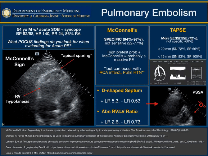
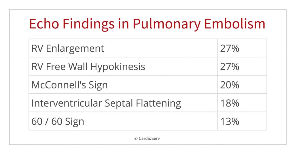
Mcconnell s sign 20.
Rv lv ratio pulmonary embolism.

Eight Pearls For The Crashing Patient With Massive Pe
Pulmonary Embolism In Echo

Moderate Acute Dilatation Of The Rv In A 55 Year Old Man With Massive Download Scientific Diagram
Imaging Of Pulmonary Embolism
Pulmonary Embolism Litfl Ccc Respiratory

Right Heart Strain Radiology Reference Article Radiopaedia Org

Echocardiography In Pulmonary Embolism The Clot Thickens Pulmonary Embolism Pulmonary Pulmonary Emboli

Pin On Structure And Function

Echocardiographic Examination Of A Patient Admitted For A Recurrent Download Scientific Diagram
Peripheral Matters Current Status Of Interventional Therapies In Acute Pulmonary Embolism American College Of Cardiology

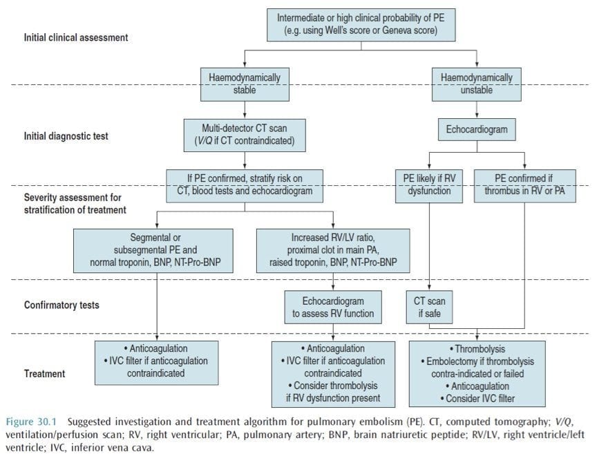
2019 Esc Guidelines For The Diagnosis And Management Of Acute Pulmonary Embolism Developed In Collaboration With The European Respiratory Society Ers European Respiratory Society

Chronic Pulmonary Embolism Diagnosis Abstract Europe Pmc

Original And Simplified Pulmonary Embolism Severity Index Download Table
Plos One Association Between Computed Tomography Obstruction Index And Mortality In Elderly Patients With Acute Pulmonary Embolism A Prospective Validation Study

Diagnostic Implications Of Computed Tomography Pulmonary Angiography In Patients With Pulmonary Embolism
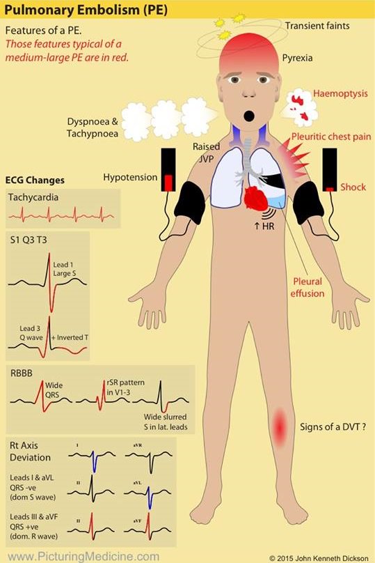
Signs And Symptoms Of Pulmonary Embolism Pe Pulmonary Embolism Signs Symptoms Acutepe Diagnosis

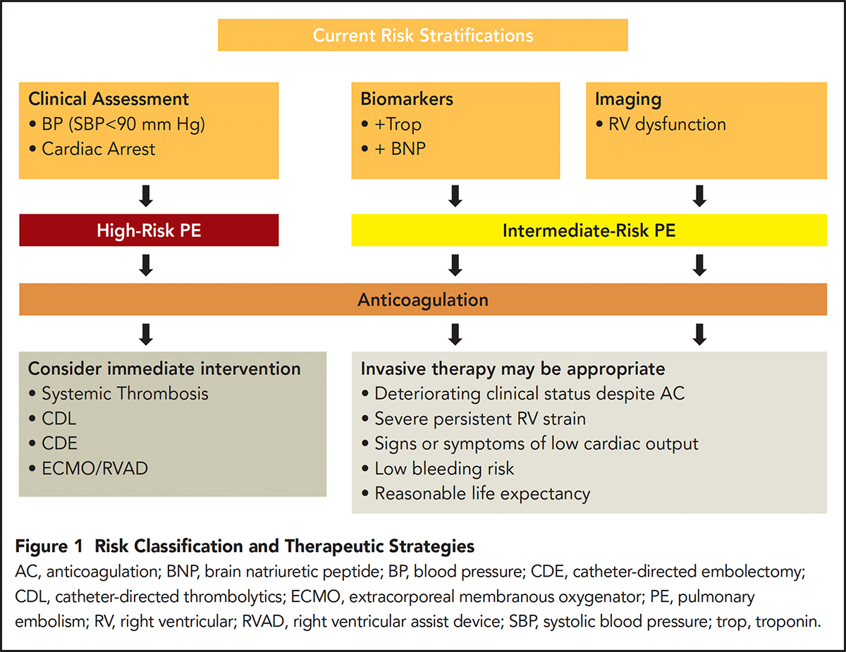
Pdf Management Of Patients With High Risk Pulmonary Embolism A Narrative Review

Submassive Massive Pe Emcrit Project
Https Encrypted Tbn0 Gstatic Com Images Q Tbn 3aand9gcsmnit Cbktx7mceehchohiwmhirjkzwqfopwqqwwqiml8g5yti Usqp Cau

A Novel Ecg Parameter For Diagnosis Of Acute Pulmonary Embolism Rs Time Rs Time In Acute Pulmonary Embolism Sciencedirect
Saddle Pulmonary Embolus Jetem

Pert Activation Pert Indicates Pulmonary Embolism Response Team Download Scientific Diagram
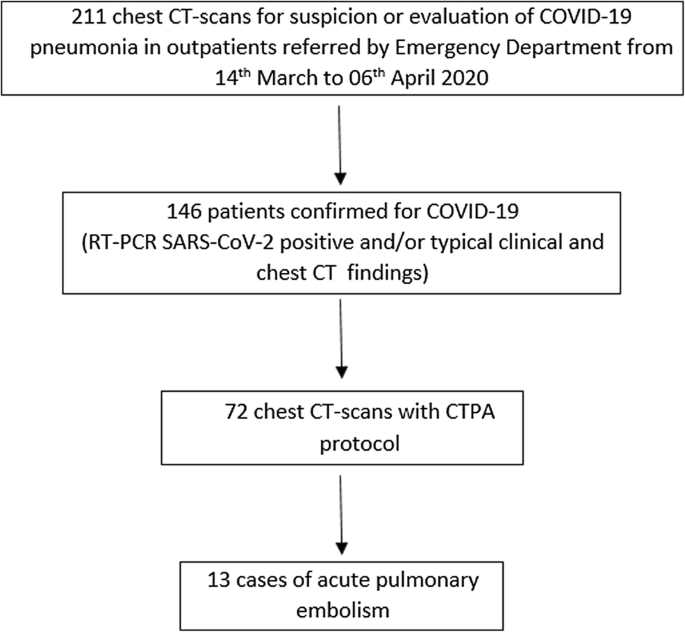
Acute Pulmonary Embolism In Non Hospitalized Covid 19 Patients Referred To Ctpa By Emergency Department Springerlink

New Horizons In Pulmonary Embolism Treatment
Source : pinterest.com
